Pros and Cons of Iron Supplementation in Children During an Illness
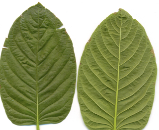
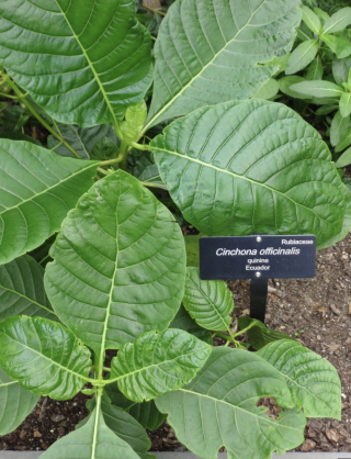
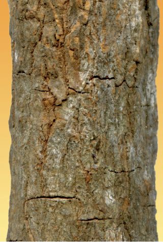
While Cinchona officinalis bark powder was once part of an effective remedy for diphtheria in the past, Cinchona officinalis doesn’t contain a lot of copper as a natural component that would increase iron levels in a safe and balanced way inside the body. But homeopathic preparations of Cinchona officinalis (sometimes called China officinalis) contains copper as an added ingredient perhaps because quinine alters iron metabolism while the extra, trace / homeopathic dose of copper improves the body’s overall use of iron and the production of healthy red blood cells nonetheless. In other words, quinine forms an alliance with iron in red blood cells such that quinine (unlike iron) makes heme toxic to pathogens. In malaria parasites that eat their host’s hemoglobin, for example, the parasite normally tries to convert the heme in hemoglobin into a non-toxic substance called hemozoin. But quinine makes a complex with the heme that makes it impossible for the parasite to neutralize the heme into hemozoin. Accumulation of heme in the parasite’s gut ultimately kills it. In sick patients who have been given quinine, ideally via hourly dosing, malaria parasites lack access to non-toxic sources of iron. This makes the malaria pathogen weak and vulnerable to other types of medicinal treatments which, in the case of malaria, often include Artemisinin (derived from Artemisia annua). Artemisinin and quinine work very well together through their different mechanisms of action against malaria.
Click here to schedule a health coaching session with us.
Artemisinin is activated by iron. Unlike artemisinin, quinine is not activated by iron. Rather, quinine binds to the iron in heme to create a toxic situation for the malaria parasite. Though diphtheria has not been studied in terms of quinine uptake, Staphylococcus bacteria have. Staphylococcus bacteria do take up quinine for the same reasons that malaria takes up quinine – they’re trying to gain access to iron that’s stored as heme in our red blood cells. Like malaria protozoa, when Staphyloccoccus bacteria seek out heme in our red blood cells in a patient treated with proper doses of quinine or Cinchona bark, the quinine kills them.Corynebacterium diphtereriae interacts differently with quinine than Staphylococcus aureus and malaria protozoa. The Corynebacterium diphtheriae bacteria, after all, is not pathogenic to humans until it is infected with a bacteriophage that then causes the diphtheria bacteria to release an exotoxin. The exotoxin primarily causes harm by preventing human cells from producing needed proteins which can lead to cell death and tissue destruction. The diphtheria toxin can damage many different types of tissues in the body, but while Staphylococcus aureus bacteria and malaria protozoa are hungry for iron from the heme in our red blood cells, some variants of diphtheria bacteria (such as Mitis, Intermedius, and Belfanti) may work to destroy red blood cells within a small, localized area of the body too. The diphtheria bacteria will completely destroy red blood cells which then become incorporated into the pseudomembrane in the throat, for example. The pseudomembrane is made up of other blood components like white blood cells and fibrin too.
Similar to Staphylococcus bacteria and malaria protozoa, the Corynebacterium diphtheriae uses heme as a source of iron particularly when the patient is iron deficient already. This explains why doctors in the 1800s and early 1900s noted that correcting iron deficiency was central to treating diphtheria. But the relationship between Corynebacterium diphtheria and iron is less straightforward than the relationships we’ve described between Staphylococcus and malaria protozoa. In a diphtheria infection, diphtheria toxin production is regulated by iron levels in the host’s infecting diphtheria bacterium. Most strains of infectious diphtheria bacteria require nicotinic acids (vitamin B3) and pantothenic acids (vitamin B5) for growth. Some also require thiamine (vitamin B1), biotin (vitamin B7), or pimelic acid (a vitamin B7 precursor). But for the optimum production of diphtheria toxin, the host should also have plenty of amino acids and be iron deficient.
Click here to buy Cinchona officinalis / Quina.
A large number of bacteria need a good source of iron from the host (usually hemoglobin) in order to cause disease. So this is odd. The diphtheria bacteria requires a good source of B vitamins and amino acids along with iron deficiency in order to produce the diphtheria toxin. Though iron deficiency optimizes the production of the diphtheria toxin, some strains of Corynebacterium diphtheria use the hemin iron from the hemoglobin-haptoglobin complex in blood plasma. So in other words, rather than destroying red blood cells themselves to obtain iron, the diphtheria pathogen taps into the body’s iron stores that exist in the liquid plasma of the blood supply.So essentially, though the diphtheria pathogen does, indeed, like to snack on iron, it doesn’t seek out and destroy human red blood cells to gain access to it. Rather, we might think of diphtheria as a vulture-pathogen that consumes the iron-leftovers in the plasma when the body has a low-level infection with Streptococcus Group A or certain Staphylococcus bacteria. Essentially, these bacteria can destroy red blood cells and leave behind heme that is sopped up by the hemoglobin-haptoglobin complex in the blood plasma. If Streptococcus or Staphylococcus colonize the body, they may slowly erode away at the iron stores to produce anemia, creating the optimal environment for a diphtheria infection.
A strep infection involving sore throat and the characteristic “white spots” can, in fact, look very similar to diphtheria and vice versa. In the 1920s, doctors often struggled to properly differentiate between strep infections / scarlet fever and diphtheria. One doctor wrote a summary of this struggle in 1927, noting specifically that, “scarlet fever often develops in patients admitted to diphtheria wards” and “in a study of 220 deaths from diphtheria, scarlet fever was reported 34 times”.
Click here to buy Artemisinin.
At that time, before vaccination became common and before these childhood diseases became less frequent, doctors knew that the simultaneous development of diphtheria and a strep infection was common. After a swab of diphtheria lesions at that time for culturing, doctors were well aware of the fact that streptococcal bacteria could be cultured too in addition to the diphtheria bacteria. Indeed, streptococcal bacteria were not the only type of bacteria found cohabiting in diphtheria lesions, but they were by far the most common.As it turns out, the streptococcal bacteria (both Group A and Group B), including Streptococcus pyogenes, the bacteria that causes strep throat and scarlet fever, produce exotoxins known as “streptolysins” that cause complete destruction of red blood cells. So, as we’ve already discussed, these bacteria can produce hemolytic anemia. The streptococcal bacteria are skilled at colonizing the body in addition to their ability to cause outright infection. In children, a colony of streptococcal bacteria can cause iron-deficiency and anemia, creating the ideal conditions for a diphtheria infection. Streptolysins can destroy red blood cells so that the streptococcal bacteria can consume most of the iron, leaving behind just a bit that ends up being captured in the hemoglobin-haptoglobin complex of the blood plasma.
Click here to do a free trial of EMDR to Release Trauma.
Other pathogens that cause red blood cell destruction, iron-deficiency, and a potentially conducive environment for diphtheria infection include:- Plasmodium spp. / Malaria
-
-
- Malaria parasites directly invade red blood cells where the parasites multiply, the red blood cell bursts, and the parasites are then released - this disease causes massive red blood cell destruction
-
- Clostridium perfringens
-
-
- produces a potent toxin (lecithinase) that destroys red blood cells in cases of severe sepsis or gangrene
-
- Streptococcal bacteria
- Streptococcus pyogenes (Group A Strep)
-
-
-
- produce exotoxins called streptolysins that causes the complete lysis of red blood cells
-
-
- Streptococcus agalactiae (Group B Strep)
-
-
-
- produce hydrogen peroxide that causes alpha-hemolysis (partial, greenish lysis)
-
-
- Staphylocccus aureus
-
-
- produces hemolysis that cause complete or partial lysis of red blood cells - the toxin, alpha-hemolysin, makes a hole in the red blood cell that slowly causes the red blood cell to leak or “lyse”
-
- Escherichia coli
-
-
- Shiga-toxin-producing E. coli (STEC) causes hemolytic uremic syndrome (HUS) that damages the lining of small blood vessels leading to mechanical fragmentation and destruction of red blood cells.
- Some E. coli strains produce hemolysins that destroy red blood cells
-
- Haemophilus influenzae
-
-
- causes immune complex-mediated hemolysis - the body’s antibodies try to kill the H. influenzae pathogen, inadvertendly causing red blood cell destruction
-
- Bartonella bacilloformis
-
-
- responsible for Carrion’s disease / Oroya fever - it invades and destroys red blood cells
-
- Babesia
-
-
- Parasites infect red blood cells, similar to malaria, ultimately causing massive red blood cell destruction
-
- Human Immunodeficiency Virus / HIV
-
-
- triggers the immune system to destroy red blood cells
-
- Epstein-Barr Virus / EBV
-
-
- produces an immune response causing the body to destroy its own red blood cells
-
- Cytomegalovirus
-
-
- produces an immune response causing the body to destroy its own red blood cells
-
- Mycoplasma pneumoniae
-
- causes cold agglutinin disease, a so-called autoimmune disease where the body’s immune system attacks red blood cells at cold temperature
Click here to subscribe to the Living Database now.
Resources:
• Linus Pauling Institute (2026)). Copper. Retrieved January 6, 2025 from https://lpi.oregonstate.edu/mic/minerals/copper#:~:text=Adequate%20copper%20nutritional%20status%20is,absorption%20in%20infants%20(24).
• Olsnes, S. and Sandvig, K. (1986). Interactions between diphtheria toxin entry and anion transport in Vero cells. II. Inhibition of anion antiport by diphtheria toxin. Retrieved January 6, 2025 from https://pubmed.ncbi.nlm.nih.gov/3944101/#:~:text=Abstract,ability%20to%20bind%20the%20toxin.
• Ferrer, P. et al. (2012). Antimalarial Iron Chelator, FBSO701, Shows Asexual and Gametocyte Plasmodium falciparum Activity and Single Oral Dose Cure in a Murine Malaria Model. Retrieved January 7, 2025 from https://journals.plos.org/plosone/article?id=10.1371/journal.pone.0037171#:~:text=Iron%20metabolism%20is%20a%20proven,severe%20cerebral%20malaria%20%5B5%5D.
• Shoji, O. (2023). Comprehensive Inorganic Chemistry III (3rd Ed): Bioinorganic Chemistry and Homogenous Biomimetic Inorganic Catalysis. Retrieved January 7, 2025 from https://www.sciencedirect.com/topics/chemistry/protoporphyrin-ix#:~:text=Abstract,molecules)%20are%20briefly%20covered%20herein.
• Kremyanskaya, M. (2025). Modulators of the Hepcidin Pathway in Polycythemia Vera and Myelofibrosis. Retrieved January 7, 2025 from https://ashpublications.org/blood/article-abstract/doi/10.1182/blood.2025028643/547823/Modulators-of-the-Hepcidin-Pathway-in-Polycythemia?redirectedFrom=fulltext
• Mabasa, V. et al. (2025). Expression of salivary hepcidin and its inducer, interleukin 6 as well as type I interferons are significantly elevated in infants with poor oral rotavirus vaccine take in South Africa. Retrieved January 7, 2025 from https://www.frontiersin.org/journals/immunology/articles/10.3389/fimmu.2025.1517893/full
• Latarissa, I. R. et al. (2021). Potential of Quinine Sulfate for COVID-19 Treatment and Its Safety Profile: Review. Retrieved January 7, 2025 from https://pmc.ncbi.nlm.nih.gov/articles/PMC8665662/
• Root-Bernstein, R. et al. (2009). Antigenic complementarity between coxsackie virus and streptococcus in the induction of rheumatic heart disease and autoimmune myocarditis. Retrieved January 19, 2026 from https://pubmed.ncbi.nlm.nih.gov/18608177/
• Patterson, M. J. (1996). Medical Microbiology, 4th Ed.: Chapter 13 Streptococcus. Retrieved January 19, 2026 from https://www.ncbi.nlm.nih.gov/sites/books/NBK7611/#:~:text=Figure%2013%2D1,not%20grow%20on%20routine%20subculture.
• Kanso, M. A. et al. (2022). Coronavirus pleomorphism. Retrieved January 19, 2026 from https://pubs.aip.org/aip/pof/article/34/6/063101/2846764/Coronavirus-pleomorphism
• Mathan, M. et al. (1975). Pleomorphic Virus-Like Particles in Human Faeces. Retrieved January 19, 2026 from https://www.sciencedirect.com/science/article/pii/S0140673675918322